Retinol in Adults Averaging 87: What a 24-Week Arm Study Measured
A small controlled trial found better wrinkle scores and tissue-marker changes with 0.4% retinol. Its strongest lesson is how to separate visible improvement, microscopic findings, and claims the experiment never tested.
Public-source editorial review. No clinician sign-off, firsthand testing, patient-image assessment or individual prescription advice claimed.
One of the more distinctive retinol experiments enrolled 36 older adults whose average age was 87. Rather than comparing facial photographs from unrelated people, researchers treated opposite arms within the same participant: one with a retinol lotion and the other with its vehicle, the lotion base without retinol. Over 24 weeks, fine-wrinkle scores improved more on the retinol-treated side. That is meaningful evidence for a defined preparation under controlled conditions—not a demonstration that every retinol product produces the same result.
The numerical result deserves particular care. A reported change of −1.64 was a decrease in score points on a 0–9 severity scale, not a 64% reduction in wrinkles. Small biopsy subgroups also showed changes in tissue markers, but those measurements were not wrinkle counts or proof of stronger, injury-resistant skin. This analysis uses the indexed primary research abstracts and the available official RENOVA labeling; it does not assume access to the studies’ complete protocols, supplements, or full reports.
Within this reading
Study facts at a glance
Population and design: The retinol trial enrolled 36 residents of two senior facilities, with a mean age of 87, in a randomized, double-blind comparison of opposite arms. Source 1
Studied exposure: Study personnel applied topical 0.4% retinol lotion or vehicle up to three times weekly for 24 weeks. This describes the experiment, not a recommended routine. Source 1
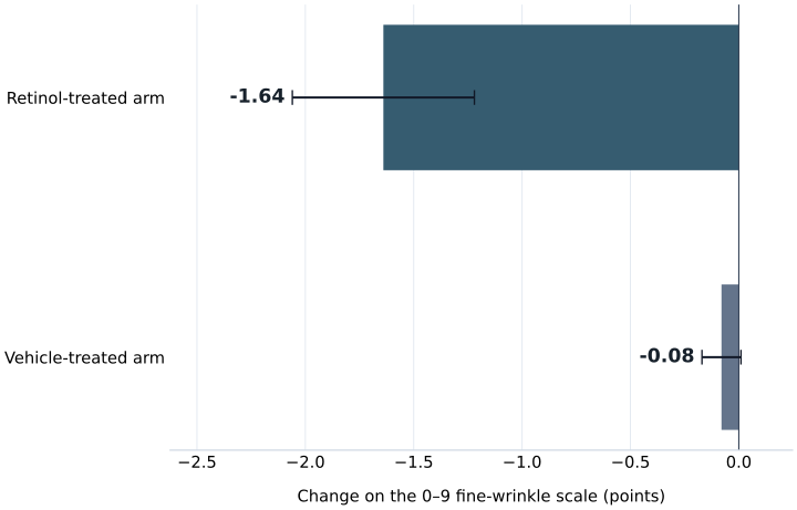
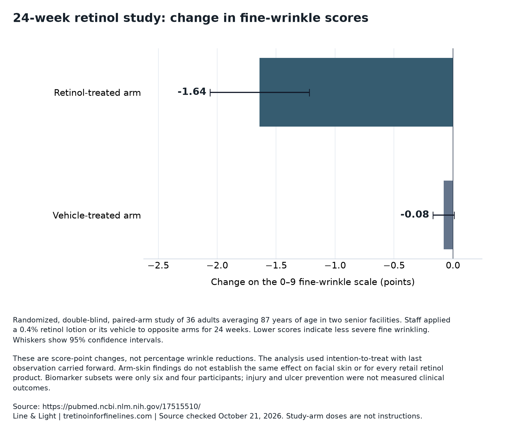
Fine-wrinkle result: Mean score change was −1.64 with retinol versus −0.08 with vehicle; the reported comparison had P<.001. These are score-point changes, not percentage reductions. Source 1
Biopsy evidence: Glycosaminoglycan expression increased in a subgroup of six, and procollagen I staining increased in a subgroup of four. The abstract gives no percentage increase for either marker. Source 1
Concentration is not an efficacy ladder: A separate 48-week facial-photoaging study found similar improvement with 0.1% and 0.025% tretinoin, but significantly more redness and scaling with the higher concentration. Source 2
Why opposite arms made a useful comparison
Kafi and colleagues studied naturally aged skin in residents of two senior facilities. Each participant contributed a retinol-treated arm and a vehicle-treated arm, with treatment side randomized. This within-person design helps control for characteristics shared by both arms, including age and many individual influences on skin appearance. It is more informative than comparing treated older adults with a separate untreated group whose skin might differ at baseline.
The study was double-blind, meaning treatment identity was concealed from relevant participants and evaluators, although the indexed abstract does not describe the detailed masking procedures. The vehicle comparator also matters: a lotion base can affect appearance independently of its active ingredient. The experiment therefore asked whether adding retinol produced a benefit beyond the base, not whether lotion was better than doing nothing.
Thirty-six participants still represent a small and unusually old population. The paired design improves the comparison’s efficiency, but it does not broaden its reach to younger facial skin. The main outcome measures combined clinical severity assessment with biochemical measurements from treated-site biopsies—two different kinds of evidence.
Read the underlying chart data: download CSV or download the source-labeled chart.
The treatment was a controlled exposure, not a retail routine
Study personnel applied 0.4% retinol lotion and its vehicle at visits, up to three times a week for 24 weeks. The route was topical: the preparation went onto arm skin. Staff application distinguishes the experiment from ordinary home use, where application quantity, missed applications, accompanying products, and treatment interruptions may vary.
The phrase up to three times weekly does not establish that every participant received exactly three applications every week. Nor does the abstract supply enough detail to reconstruct each person’s cumulative exposure. The concentration, formulation, application setting, body site, and duration together define the tested intervention; concentration alone does not.
Retinol is not tretinoin, which is all-trans-retinoic acid. Their percentages cannot be treated as equivalent units of clinical potency. Likewise, evidence for this retinol lotion is not automatic evidence for other over-the-counter lotions, compounded preparations, oral products, or differently formulated creams. The preparation-comparison guide provides a useful framework for keeping product identity separate from an ingredient-level promise.
Reading the wrinkle score without turning it into a percentage
Clinical appearance was assessed on a semiquantitative scale ranging from 0, no severity, to 9, greatest severity. Semiquantitative means that observations are assigned numerical grades rather than measured as an exact physical quantity such as wrinkle depth in millimeters. Lower scores were better, so negative changes indicated improvement.
At 24 weeks, the reported fine-wrinkle change was −1.64 with retinol and −0.08 with vehicle. Subtracting those changes gives an editorially calculated difference of 1.56 score points favoring retinol. That calculation describes separation between average score changes. It does not tell us how many wrinkles disappeared, what fraction of participants noticed improvement, or how deep an individual line became.
A percentage reduction would require an appropriate denominator and a scale that supports that interpretation. Neither the digits 64 nor division by the nine-point range supplies one. Baseline severity and the clinical meaning of a one-point step also matter. Our guide to fine lines versus deeper wrinkles explains why an improvement in one appearance category should not be stretched into a claim about another.
What the retinol measurements establish
| Measurement | Reported result | Correct interpretation |
|---|---|---|
| Fine-wrinkle score change | Retinol −1.64; vehicle −0.08 | Average changes in severity-score points, not percentages |
| 95% confidence intervals | Retinol −2.06 to −1.22; vehicle −0.17 to 0.01 | Uncertainty intervals for each reported change, not an interval for their difference |
| Glycosaminoglycan expression | n=6; P=.02 | A small-subgroup tissue-marker finding; magnitude not supplied |
| Procollagen I immunostaining | n=4; P=.049 | A small-subgroup staining finding, not a reported percent increase in collagen |
Strong statistical separation still leaves clinical questions
The retinol change had a 95% confidence interval from −2.06 to −1.22; the vehicle change ranged from −0.17 to 0.01. These intervals describe uncertainty around the separate changes. The vehicle interval includes zero, whereas the retinol interval does not. The report also gives P<.001 for the comparison, supporting a difference under the statistical analysis used.
The P value is not the probability that retinol works for a particular person. Nor does statistical significance establish how noticeable a 1.56-point average separation would be in everyday life. The abstract does not provide a patient-rated importance threshold, the distribution of individual responses, or a percentage reaching a prespecified visible-improvement target.
The available estimate is tied to 24 weeks. It does not establish the earliest reliable onset, whether benefit continued increasing afterward, or how long improvement remained after treatment ended. These are distinct time-course questions. A fine-line photograph can illustrate appearance, but it cannot replace a defined scoring system, a comparator, and an explicit assessment time.
Missing-data handling is not evidence of complete follow-up
The investigators reported an intent-to-treat analysis using last observation carried forward. Intent-to-treat generally aims to preserve the original treatment assignment in the analysis. Last observation carried forward fills a missing later measurement with an earlier available measurement, effectively assuming no further change after that observation.
That approach can influence the result if participants with missing measurements would otherwise have improved or worsened. It is not necessarily conservative in every setting. Its effect depends on when data were missing, why they were missing, and how skin scores would have developed afterward.
Importantly, mentioning this method does not mean there was no attrition. The indexed abstract does not provide the completion count, missing-data pattern, or alternative analyses testing different assumptions. Those omissions limit assessment of robustness without erasing the randomized comparison. They identify what a fuller appraisal would need: retention, reasons for missing visits, and sensitivity of the estimate to the imputation method.
Microscopy supported a biological response, not a collagen percentage
The biopsy results came from much smaller groups than the clinical analysis. Glycosaminoglycan expression increased relative to vehicle in six participants, with P=.02. Procollagen I immunostaining increased in four, with P=.049. These sample sizes must remain attached to the findings; neither tissue result represents a biopsy measurement in all 36 participants.
Glycosaminoglycans are tissue components associated with water retention. Procollagen I is a precursor associated with collagen production, and immunostaining uses antibodies to reveal a target in tissue. The findings are consistent with a biological response that could help explain smoother appearance. They do not quantify total mature collagen, its organization, or mechanical skin strength.
The abstract reports significance levels but not effect sizes or confidence intervals for these markers. Consequently, it cannot support a claim of a particular percentage increase in collagen or hydration. The RENOVA label makes a related distinction for tretinoin: increased procollagen staining has not been clearly linked to collagen I levels or observed clinical effects. Appearance and tissue markers can support a coherent hypothesis without proving the entire causal chain.
Injury resistance remained a hypothesis—and safety counts remain unavailable
The retinol authors proposed that increased skin-matrix synthesis might make aged skin better able to withstand injury and ulcer formation. That is a hypothesis extending beyond the measured outcomes. The abstract does not report an ulcer-prevention endpoint, injury rates, wound healing, or a mechanical-resilience test. Better wrinkle scores cannot stand in for fewer clinical ulcers.
The same boundary applies to tolerability. The indexed retinol abstract does not provide complete adverse-event counts, irritation severity, or treatment-discontinuation counts. It therefore cannot establish zero irritation, universal tolerability, or a numerical benefit-to-harm balance. Staff application in a supervised study is relevant context, but it does not fill in the missing safety results.
Related tretinoin evidence documents real local adverse effects. In the concentration trial, redness and scaling were significantly greater with 0.1% than with 0.025%. RENOVA labeling describes peeling, dryness, burning, stinging, redness, and itching. Those observations identify concerns with related treatments; their frequencies should not be assigned to the retinol lotion studied in these older adults.
The 48-week tretinoin trial challenges stronger-is-better reasoning
A separate double-blind trial reported 99 photoaged patients who completed 48 weeks: 32 used 0.1% tretinoin cream, 35 used 0.025%, and 32 used vehicle. The preparations were applied topically once daily. Both active groups improved facial photoaging relative to vehicle, with no clinically or statistically significant efficacy difference reported between the concentrations.
Tissue findings followed a similar pattern. Epidermal thickness increased by 30% and 28% in the higher- and lower-concentration groups, compared with an 11% decrease with vehicle. Vascularity increased by 100% and 89%, compared with a 9% decrease. These are percentages for particular tissue measurements—not percentages of wrinkles removed. Irritation was greater with the higher concentration despite similar reported clinical improvement.
This separation argues against treating irritation as a necessary measure of efficacy. It does not prove that all strengths are interchangeable or that the study formally established equivalence. The 48-week concentration-trial analysis examines that distinction. Different drug, population, body site, and follow-up prevent an efficacy ranking against the 24-week retinol arm study.
Three experiments answering different questions
| Study | Population and exposure | Main interpretive boundary |
|---|---|---|
| 24-week retinol study | 36 older adults; mean age 87; opposite arms; 0.4% retinol versus vehicle | Naturally aged arm-skin findings do not establish facial results for all retinol products |
| 48-week concentration study | 99 completers with photoaging; facial 0.1% or 0.025% tretinoin versus vehicle | Similar reported efficacy does not provide a retinol-to-tretinoin conversion |
| Two-year tretinoin study | 204 subjects; moderate-to-severe facial photodamage; 0.05% emollient cream versus vehicle | Longer-term findings concern this preparation and population, not every retinoid |
Two-year findings and current labeling answer different questions
The longer trial enrolled 204 subjects with moderate-to-severe facial photodamage. Tretinoin emollient cream 0.05% or vehicle was applied to the face once daily for up to two years. The abstract reports significantly greater improvement with tretinoin across several clinical assessments, including fine and coarse wrinkling and overall photodamage, but does not give numerical effect sizes or confidence intervals for those benefits.
Histology showed no increase relative to placebo in keratinocytic or melanocytic atypia, dermal elastosis, or adverse stratum-corneum findings. A procollagen-synthesis marker increased at month 12. These are specific reassuring observations, not proof of an absence of all adverse effects; the abstract does not supply a complete local-reaction accounting.
Current RENOVA 0.02% labeling concerns a different exact cream. It indicates adjunctive mitigation of fine facial wrinkles with comprehensive skin care and sunlight avoidance, not wrinkle elimination or reversal of photoaging. It also reports that about 4% discontinued because of adverse reactions. Its discussion of ages 65–71 does not establish effectiveness above 71, much less at an average age of 87. The RENOVA indication guide keeps those product-specific limits together.
Translating the experiment into evidence questions
The strongest practical reading is to ask whether a claim preserves the experiment’s identity. Does it name the tested formulation, arm location, 24-week duration, score-based outcome, and older population? Does it distinguish a clinical score from a biopsy marker? A statement that simply says retinol rebuilds skin leaves out most of the information needed to judge what was demonstrated.
Editorial interpretation: when considering a paid service, the scientific result and the service contract are separate documents. A trial cannot establish a provider’s formulation, follow-up arrangements, renewal terms, or outcomes. Readers can use the Dermatica review and Nurx review to check documentary terms, without assuming either service reproduces this intervention or offers care for this study population.
Funding and sponsorship also belong in an evidence appraisal. The accessible research abstracts do not identify sponsors, so sponsor influence cannot be assessed here. The official label identifies Bausch Health entities as RENOVA’s distributor and manufacturer; that does not identify the retinol trial’s sponsor. The unresolved scientific questions remain concrete: response variability, durability, complete tolerability, and whether marker changes predict meaningful tissue function.
Questions about the evidence
Did retinol reduce wrinkles by 64%?
No. The reported −1.64 was an average change in severity-score points on a 0–9 scale. Vehicle-treated skin changed by −0.08 points. Neither number is a percentage reduction, a wrinkle count, or a measurement of wrinkle depth.
Does the study show that retinol works on an 87-year-old face?
It provides evidence for naturally aged arm skin in a group whose mean age was 87. It did not test facial application. A mean age also does not describe every participant’s age or establish an individual response. Facial results require evidence appropriate to that site and preparation.
Did all 36 participants have increased collagen?
No such result is reported. Procollagen I staining was assessed in a subgroup of four, and glycosaminoglycan expression in six. The abstract reports statistical comparisons but no percentage increase in total collagen. A precursor-marker finding is not the same measurement as mature collagen content.
Can 0.4% retinol be converted into an equivalent tretinoin strength?
These studies provide no conversion. They used different ingredients, formulations, populations, treatment schedules, and skin sites. Even within the tretinoin concentration trial, the higher percentage produced more irritation without a reported efficacy advantage, illustrating why concentration alone is an inadequate clinical ranking.
Did the trial establish ulcer prevention or long-term safety?
No. Injury and ulcer resilience were proposed implications, not reported clinical endpoints. Follow-up lasted 24 weeks, and the indexed abstract does not give complete adverse-event counts. Separate long-term tretinoin findings cannot supply missing safety or ulcer-prevention results for this retinol lotion.
What this evidence supports
The 24-week trial supports a specific conclusion: staff-applied 0.4% retinol lotion improved graded fine wrinkling of naturally aged arm skin more than its vehicle in a small, very old population. Its paired, randomized design strengthens that comparison. The biopsy findings add biological plausibility, but their tiny subgroups and unreported effect magnitudes limit interpretation. Neither percentage wrinkle removal, prevention of ulcers, equivalence to tretinoin, nor performance of unrelated retail products follows from these results. For Line & Light’s measurement perspective, the central distinction is between a visible severity score, a tissue marker, and a functional clinical outcome—three endpoints that should never be substituted for one another.
Research access and limitations
Research analysis is based on indexed primary-report abstracts, alongside official current RENOVA labeling retrieved in full. Complete trial protocols, participant-level data, supplements, funding disclosures, and full adverse-event tables were not available for this analysis. Study exposure descriptions are not application instructions, and trial findings are not current prescribing information.
The records behind this article
- Improvement of naturally aged skin with vitamin A (retinol). ↗Indexed original research abstract · Accessed 2026-10-21
- Two concentrations of topical tretinoin (retinoic acid) cause similar improvement of photoaging but different degrees of irritation. A double-blind, vehicle-controlled comparison of 0.1% and 0.025% tretinoin creams. ↗Indexed original research abstract · Accessed 2026-10-21
- RENOVA tretinoin cream 0.02% prescribing information ↗FDA labeling via DailyMed; record updated February 24, 2026; internal revision November 2025 · Accessed 2026-10-21
- Long-term efficacy and safety of tretinoin emollient cream 0.05% in the treatment of photodamaged facial skin: a two-year, randomized, placebo-controlled trial. ↗Indexed original research abstract · Accessed 2026-10-21